

Retatrutide and Liver Fat: Which Tests Actually Show Progress?
Everyday guidesPublished 4 min readhow to

Compare liver-fat scans, ALT, AST and fibrosis tests, with a worksheet for reading retatrutide-related results.
Illustrative licensed stock photograph. · Photo: Vitaly Gariev · pexels.com
The short answer
MRI liver-fat measurements, liver enzymes and fibrosis assessments answer different questions. Keep the same test and units when comparing progress.
Table of Contents
- What the retatrutide liver study actually measured
- Match the result to the question
- Read a percentage without turning it into a cure rate
- Make results from different clinics usable
- Four questions to take to the appointment
- Sources
- Explore this topic
Retatrutide liver-fat headlines usually describe a change measured by a particular MRI technique. A lower ALT blood result, a smaller waist and a lower liver-fat percentage are different findings. To understand progress, compare the same test over time and ask separately about fat, liver injury and scarring.
That distinction matters when a clinic package offers a long list of blood tests. More results do not necessarily answer your question better. Start with what you are trying to find out: whether fat is present, whether its amount has changed, or whether there is evidence of more serious liver disease.
What the retatrutide liver study actually measured
The 2024 randomized liver substudy included 98 people with at least 10% liver fat on MRI-PDFF. Its main endpoint was the relative change in liver fat after 24 weeks. Across the retatrutide groups, the mean reductions ranged from 42.9% to 82.4%, compared with a 0.3% increase with placebo. These were research-group results, not a forecast for an individual.
MRI-PDFF estimates the fraction of liver tissue signal attributable to fat. The study did not establish that the same percentage of fibrosis had disappeared. Blood markers also did not all move together: ALT, AST, FIB-4 and ELF did not change consistently versus placebo. This is one reason a liver-fat headline should not become a claim that every aspect of liver health has improved.
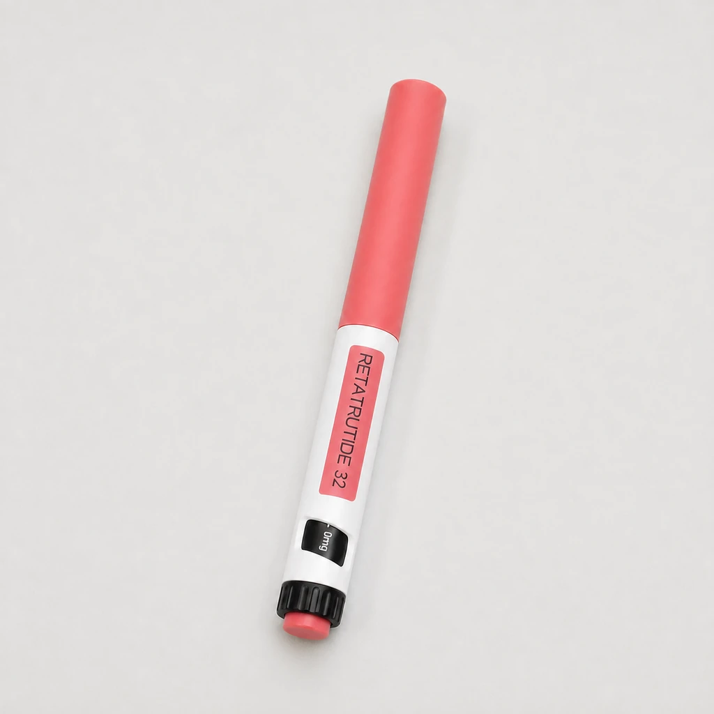
Retatrutide remains investigational as of this article's evidence check. Lilly's current development information does not establish the identity, safety or effect of products offered outside its trials.
Match the result to the question
The NIDDK explanation of liver assessment distinguishes blood tests, imaging and, in selected circumstances, biopsy. For a practical discussion, keep these categories separate:
| Result on the report | What it helps investigate | What you should not infer from it alone |
|---|---|---|
| ALT and AST | Liver-enzyme abnormalities and possible causes | An exact liver-fat percentage or a fibrosis stage |
| Ultrasound description of fatty liver | Whether the scan shows features of fat accumulation | A directly comparable numerical MRI-PDFF result |
| MRI-PDFF percentage | The measured liver-fat fraction | That inflammation or scarring has resolved |
| Elastography or liver-stiffness result | Assessment of possible fibrosis in clinical context | That a change is necessarily the same as fat loss |
| A score such as FIB-4 | A clinician's assessment of fibrosis risk using relevant inputs | A diagnosis made from an isolated online calculation |
The useful question is therefore specific: “Which result will tell us whether the concern identified at my first appointment has changed?” It is more informative than asking for a generic “liver improvement test.”
Read a percentage without turning it into a cure rate
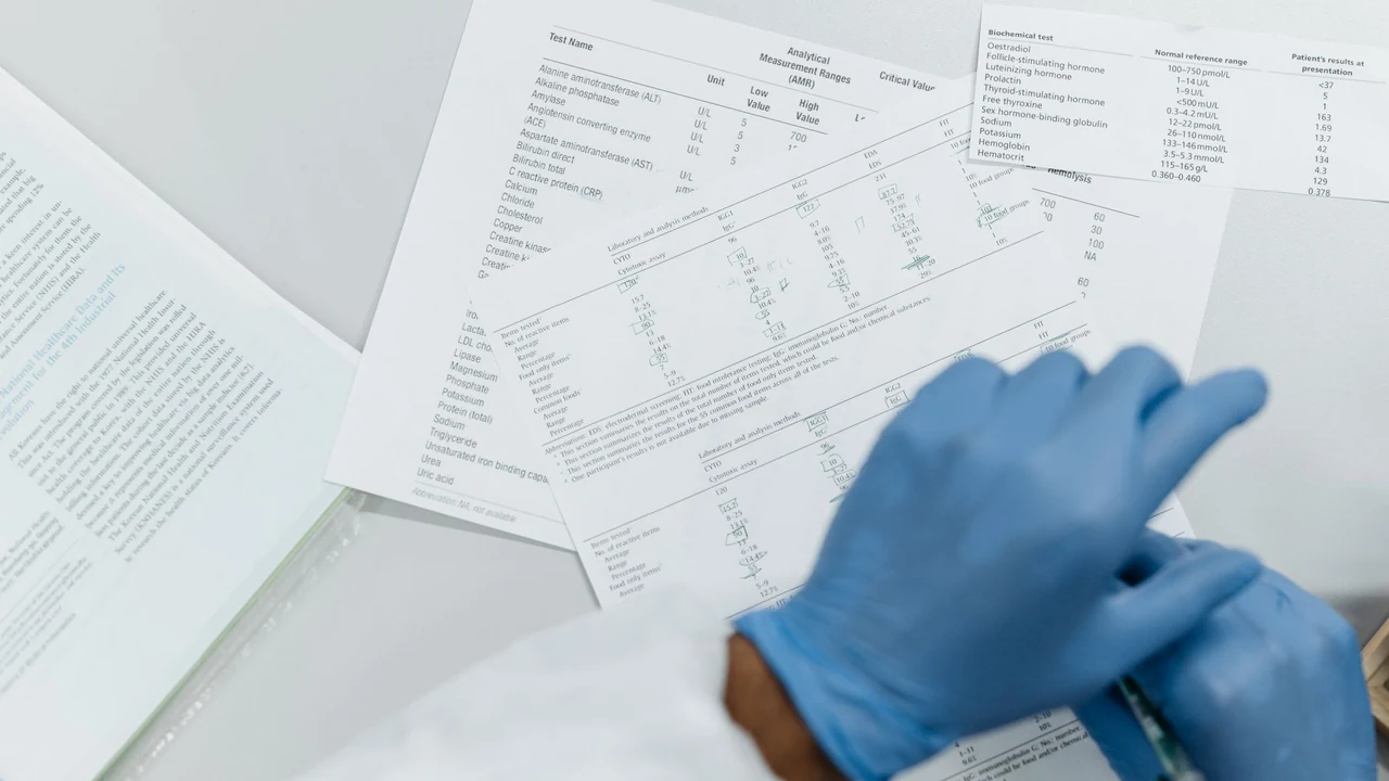
Illustrative licensed stock photograph. · Photo: Pavel Danilyuk · pexels.com
View full size ↗Here is a hypothetical calculation, not a patient result. If MRI-PDFF falls from 20% to 8%, the absolute difference is 12 percentage points. The relative reduction is 60%: divide the 12-point change by the starting 20%.
Those statements describe the same numerical change. Neither says that “60% of liver disease is cured.” They also cannot be compared directly with an ALT result falling from 80 to 40, because that is a different measurement with different units.
When you save a result, keep the original number and unit. Writing only “down 60%” removes the information needed to understand what changed. A before-and-after screenshot that cuts off the test name is particularly easy to misread.
Make results from different clinics usable
If you live between Bali and another country, your follow-up folder may contain results from several laboratories. A little organization makes the next consultation much more productive.
Create a one-page table with the date, test name, numerical result, unit, laboratory reference range and the reason the test was ordered. Attach the full imaging report, including the method and the radiologist's conclusion. Record important changes in prescribed medicines, weight and alcohol intake alongside the dates, without deciding which change caused a result.
If one report says “fatty infiltration” and the next gives a percentage, leave them as separate entries. Ask the clinician whether they can be compared. Do not invent a numerical starting point from a descriptive ultrasound label.
This also helps avoid repeating an expensive scan simply because the original report is unavailable. Bring the document itself, not only your recollection that the result was “high.”
Four questions to take to the appointment
- What is my current diagnosis, and which evidence supports it?
- Are we monitoring liver fat, possible injury, fibrosis risk, or more than one of these?
- Which test should be repeated, under what conditions, and when would the result change the plan?
- Are there other possible contributors that need assessment rather than being attributed to body weight or a peptide?
You do not need to purchase every test in the table. The point is to get a defined follow-up plan with a reason for each test. A result that would not change any decision may not answer the question you came to solve.
For the broader picture, keep a comparable weight record and discuss any existing HbA1c results separately. Neither substitutes for a liver assessment. The most useful next step is a folder of comparable evidence and a clear question, not a target percentage borrowed from a trial headline.
Instructions & useful references
- Retatrutide liver-fat randomized substudywww.nature.com · MRI-PDFF assessed liver fat in a 98-participant substudy. Fat changes are distinct from fibrosis, inflammation or clinical cure; liver blood tests did not consistently distinguish treatment groups.
- Retatrutide development statuswww.lilly.com · Current developer page checked October 11: retatrutide remains investigational; phase 3 results exist. No retail-product equivalence or approved personal regimen is inferred.
- Diagnosing fatty liver diseasewww.niddk.nih.gov · Blood tests, imaging and selected fibrosis assessment answer different diagnostic questions; ALT is not a direct liver-fat percentage.
Sources checked on October 11, 2026. This guide provides general information, with practical planning suggestions. It is not an individual treatment plan.